Behavioral effects of optogenetically induced myelination in mice
Myelination, the ‘ensheathment’ of neurons, is essential to the functioning of the central and peripheral nervous systems. So it is not surprising that problems with myelination can lead to a number of crippling diseases. Known examples include multiple sclerosis and other neurodegenerative autoimmune diseases.
Mature neurons augmenting neural circuits
In the central nervous system, oligodendrocytes produce myelin, an insulating material, which wraps around nerve axons in order to facilitate fast conduction of neural impulses. This is an especially important process during infancy, and therefore often investigated at this stage. However, how the modulation of myelination takes place after infancy is still largely unclear. Erin Gibson, David Purger, and their colleagues at the Stanford University School of Medicine (CA, USA) recently looked into the intriguing idea that postnatal myelination may be modulated by mature neurons to augment active neural circuits.
Behavioral output of myelination
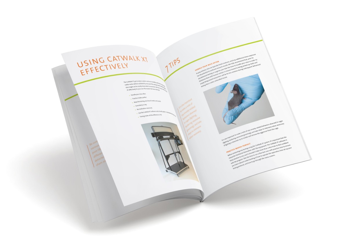
Gibson and Purger investigated how neuronal activity affected the regulation of myelin-forming cells in vivo using optogenetic stimulation of the premotor cortex in mice. They then measured behavioral output with the CatWalk XT system. CatWalk XT gait analysis is based on the voluntary movement of the animal across a glass floor. Light is reflected where paws touch the floor, allowing the camera underneath to capture actual footprints. Using these images, the software calculates parameters that are derived from individual footfalls and the time and distance relationships between footfalls.
Parameters
Three parameters were used in this study: swing speed (as a difference between the left and right forepaw), stride length (distance between successive paw placements), and paw intensity (degree of contact with the glass plate).
Myelination improves motor function
Four weeks after unilateral optogenetic stimulation, researchers expected to find improved motor behavioral performance of the correlating (in this case left) forelimb. Indeed, they found swing speed to be increased, with no change in either stride length and paw intensity. This was not caused by asymmetrical muscle development, as muscle mass and fiber diameter were unaffected. By pharmaceutically blocking oligodendrogenesis, myelination was prevented and the animals’ gait was also not affected, thus supporting the necessity of myelination for measured motor improvement.
Further research
This study shows that active neurons influence the process of myelination. Now, further research might unravel exactly what happens at the neuron level: is new myelin produced, or do existing sheets get bigger or thicker? These insights may prove useful in treating demyelinating disorders.

Gibson, E.M.; Purger, D.; Mount, C.W.; Goldstein, A.K.; Lin, G.L.; Wood, L.S.; Inema, I.; Miller, S.E.; Bieri, G.; Zuchero, J.B.; Barres, B.A.; Woo, P.J.; Vogel, H.; Monje, M. (2015). Neuronal activity promotes oligodendrogenesis and adaptive myelination in the mammalian brain. Science, 344(6183), 1252304.
Get the latest blog posts delivered to your inbox - every 15th of the month
more
What a print can tell
So what can one footprint tell you? Well, it could tell you a lot. Simply putting the paw in ink and studying the print left behind is one way to go about it, but there are far more sophisticated ways of footprint analysis.
In utero alcohol exposure, the effects on brain and behavior
About 10% of women worldwide drink during their pregnancy. This could cause the fetus to suffer from fetal alcohol syndrome, which can lead altered tissue structures in the brain to motor deficits.