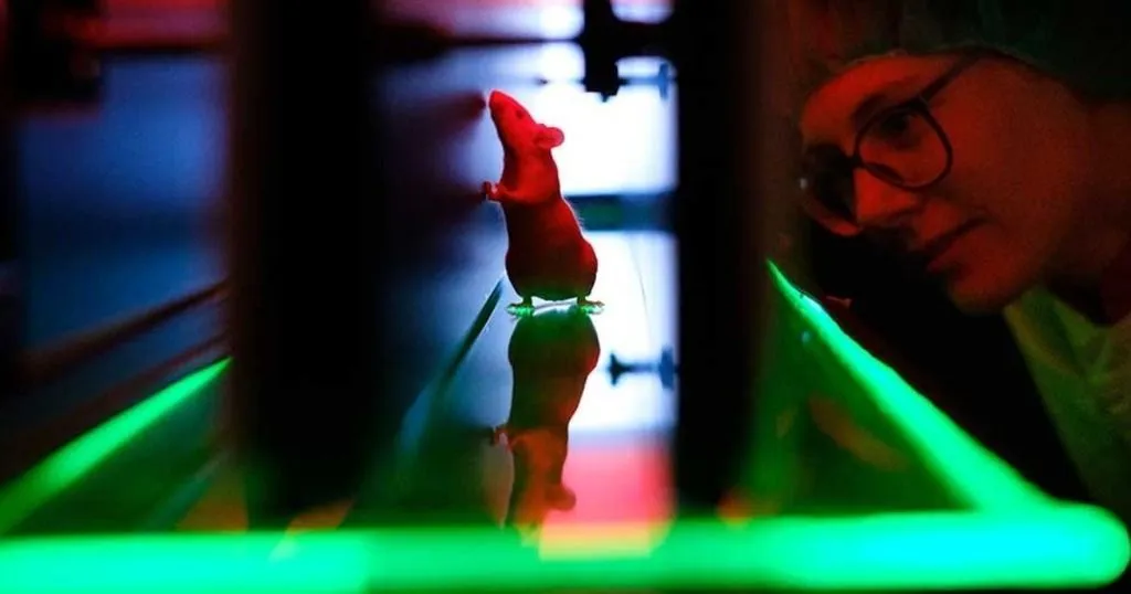
Quantifying locomotion in rats using CatWalk XT
Gait analysis is a powerful tool in evaluating behavioral and physiological changes in clinical and pre-clinical rodent models.
Read More arrow_forwardAutomated gait analysis allows for accurate assessment of several behavioral endpoints when you can’t ask your animal to please walk in a straight line.
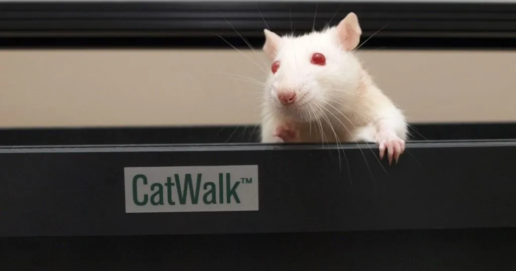
Last week I wrote about the value of a print. A footprint, that is. With CatWalk XT, you can extract a lot of information from just one footprint. In this post, I am taking it a step further by talking about the relationship between prints.
In the study of many different disorders that affect the nervous system, muscles, or bones, it is important to know how the animal walks. Does it have a regular gait, following a normal pattern of footsteps? Or can we detect a lack of coordination, or ataxia? Those are important behavioral observations in many studies. So how can you detect them: by looking at the relation between prints.
Clinicians are trained to visually observe the gait of their patients to detect abnormalities. They can ask their patients to walk a certain way or direction, to watch their stability and balance. But what if your patient is a small rodent?
Small rodents are often used in research on a wide scale of disorders that cause abnormalities in gait. Automated gait analysis allows for accurate assessment of several parameters (behavioral endpoints) when you can't ask your animal to please walk in a straight line.
In many disease models, the regularity with which the animal walks is important. For example, animal models of ataxia [1], arthritis [5,9], stroke [4], and spinal cord injury [2,3,7] have a lower regularity.

So how can you detect irregularity automatically? By using gait analysis software that recognizes 'normal' footfall patterns, and detects deviations from this pattern.
Solutions such as CatWalk XT recognizes several normal gait patterns, including: left front, right front, left hind, right hind. This pattern is described in literature as cruciate. If during the run, the animal places one of its paws outside the pattern, that paw is labeled as "not part of a pattern". The number of irregular paw placements relative to the regular paw placements is represented in the regularity index.
The base of support is the distance between either the two hind paws or the two front paws. In spinal cord injury, animals seem to have trouble keeping balance, causing them to place their hind paws wider apart than normal [3,6]. Tail and abdomen drags are not uncommon [3]. In contrast, Parkinson's models are reported to have a smaller base of support in their front paws [10], placing them closer together than non-affected controls. This is similar to the posture human Parkinson's patients show; they also keep their feet closer together and show small movements with a hunched over posture.

Stride length is the distance between the successive placement of the same paw - in other words, the distance one paw travels during one step. Again, similar to human patients, Parkinson's model animals are reported to take smaller steps [10], which can be confirmed with smaller stride lengths found with CatWalk XT automatic gait analysis. There are also reports of a decreased stride length in ataxic animals [1] and spinal cord injury models [3,8].
Curious to find out what more you can measure with automated gait analysis? Sign up to be one of the first to read the last blog in this series!
RESOURCES
Find out how CatWalk XT is used in a wide range of studies and how it can elevate your research!
Other blogs in this series are now online:
Or download the free white paper!
Of course, you can always go to www.noldus.com/catwalk-xt for more information on automated gait analysis.
Gait analysis is a powerful tool in evaluating behavioral and physiological changes in clinical and pre-clinical rodent models.
Read More arrow_forward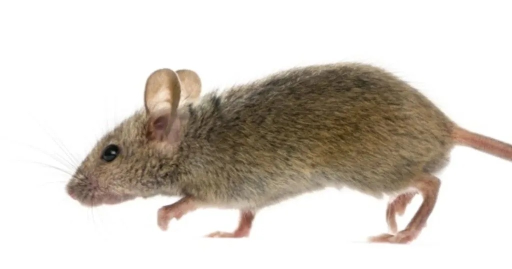
Researchers from the University of Bochum achieved significant scientific results by making mice walk again after a complete spinal cord crush! Read more about it in this blog post.
Read More arrow_forward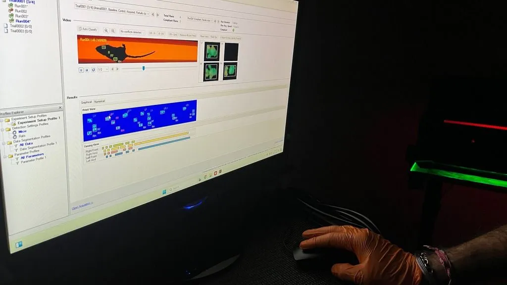
We've compiled a list of the most asked questions by CatWalk XT users. For this blog we have asked our best trainers to answer these questions. Whether you are a beginner or a seasoned user, there is something here for everyone.
Read More arrow_forwardWe'll get back to you shortly.
Please correct the following errors: