Rotator cuff injuries are quite common and have significant consequences. Almost half of the people over the age of 60 have suffered a rotator cuff tear, causing pain and decreased usability of the shoulder.
These limitations also seriously impact quality of life, and if the tendon has torn completely, it needs to be repaired surgically. The tears often heal poorly, and overall further research is warranted to improve surgical techniques.
Suture anchors vs transosseous surgery
In rats, the most commonly used surgical technique in rotator cuff tear research, the transosseous bone tunnel technique, is not the primary technique used on humans. A group of researchers from the Chinese University of Hong Kong aim to develop a new rotator cuff repair model, based on suture anchors. Suture anchors are used in the actual human surgeries that patients receive nowadays.
In the current study, a version of the suture anchors is developed that is suitable for use on rats and can be compared to the transosseous bone tunnel technique. This is especially important since surgeries that are effective on animals are often not as effective on humans, probably due to physiological differences.
An animal model on rotator cuff injuries
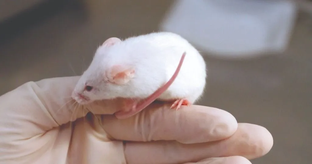
For the study, sixty male adult Sprague-Dawley rats were selected and randomly assigned to either the transosseous or the suture anchor surgery group (n = 30 per group).
Creating the rotator cuff injuries
All rats received surgical rotator cuff injuries: the supraspinatus (SS) tendon in the right shoulder was found and transected, and the rats were allowed to recover for 24 hours. After four weeks of recovery, the two respective repair surgeries were carried out.
The techniques of transosseous surgery and suture anchors
For the transosseous bone tunnel technique, the SS tendon was stitched onto the bone using holes drilled all the way through the humeral head at an angle. For the suture anchors technique, custom-made anchors were developed, since all commercially available versions were too big.
Through weaving two pieces of suture together, the scientists created a small anchor which was then pushed into a pre-drilled hole, thus attaching it to the bone. The SS tendon was then stitched with the lengths of suture sticking out from the anchor. (For images of both techniques, please take a look at the original article, referred to below.)
RESOURCES
Read more about CatWalk XT
Find out how CatWalk XT is used in a wide range of studies and how it can elevate your research!
-
check_circle
Free white papers and case studies
-
check_circle
Customer success stories
-
check_circle
Recent blog posts
Resources
arrow_forward
Gait analysis
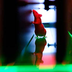
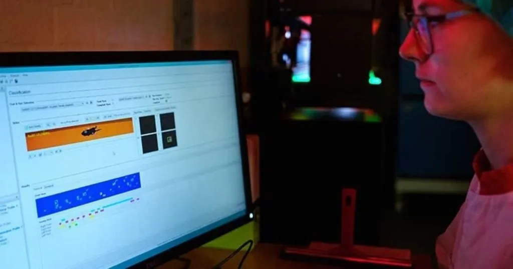
The rats underwent various tests, including gait analysis. During 2-week, 4-week, 6-week and 8-week post-repair, a downhill walking gait analysis was carried out and analyzed using Noldus' CatWalk XT.
Walking tests
Each rat could walk back and forth freely, starting from the right side of the walkway which was tilted up at 10°. The experiment stopped once the rat had finished two complete walks with a speed variation of less than 30%.
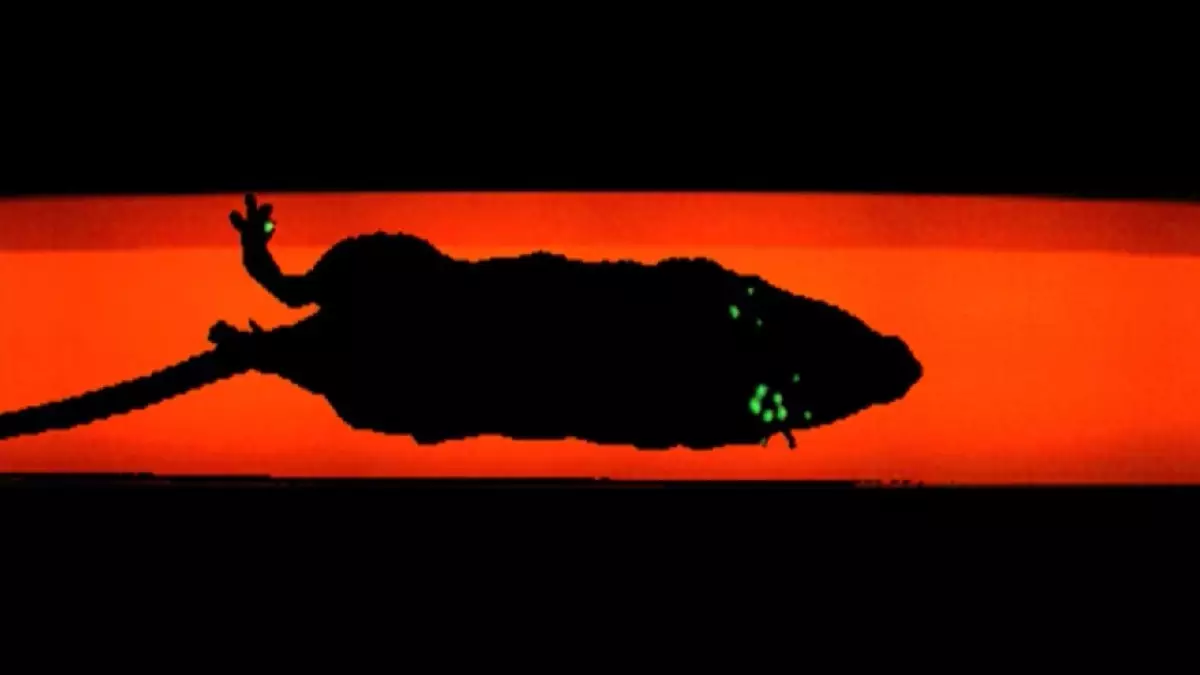
At a predetermined 10 x 60 cm area, the detection system automatically recorded the pawprint images, using a digital camera set at 60 cm below the walkway. Pawprint area, swing duration of a forelimb, max intensity and stride length were calculated automatically.
Initial recovery of rotator cuff injuries
At 4-weeks post-tear, the rats' gait had completely recovered, contrary to humans, whose shoulder functionality kept decreasing after injury. Once the tear had been repaired, the rats' gait deteriorated again for 2 weeks, and after 4 weeks returned to normal. This fast recovery is probably caused by the natural healing capacity of rats combined with the fact that rats have to put weight on their shoulder where humans do not.
Biomechanical tests
Next to gait analysis, the repaired tendons underwent various pulling strength tests. Every two weeks, shoulder samples were taken from the rats and tested to see how well the connection between bone and tendon held.
After the rats had fully healed from either surgery, the researchers found that the new tissue connecting the severed tendon to the bone was of a different structure than before surgery. Most re-tears caused by the pulling strength test took place in this soft scar tissue, indicating a lower strength of said tissue.
Healing of suture anchors vs transosseous repair
Both techniques caused the torn tendon to be fixed in place properly right after surgery. After 4 weeks, the suture anchors showed better tendon healing and decreased bone healing than transosseous repair, though after total recovery the two techniques showed comparable functional recovery.
It is possible that the initial better healing of the suture anchors is caused by compression of the tendon onto the bone. This is compensated by lesser bone damage from the drilled holes in transosseous repair, causing less swelling compared to the suture anchors.
An animal model on rotator cuff injuries
Although the suture anchors did not promote better healing compared to transosseous surgery, the Hong Kong researchers successfully developed the novel suture anchor surgery for rats. This means that future research in this topic has an increased clinical relevance and is easier and more effective to carry out.
References
Liu, Y., Fu, S. C., Yao, S. Y., Chen, X. D., & Yung, P. S. H. (2022). Application of suture anchors for a clinically relevant rat model of rotator cuff tear. Journal of Tissue Engineering and Regenerative Medicine.