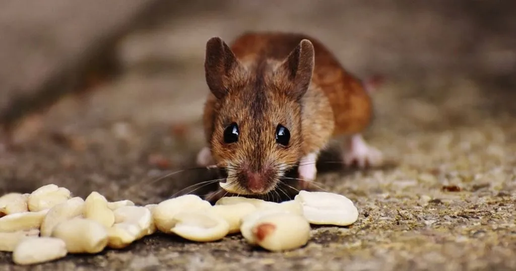
Craving a certain snack, getting it from the fridge or at the store, then the joy of eating it. It's a familiar ritual for most of us. Scientist say we are performing appetitive and consummatory behavior, and the part of our brain that regulates these behaviors is the lateral hypothalamus (LH). You can imagine that this is an important element in the investigation of, for example, addiction and eating disorders. The problem is that the LH seems to be a mosaic of different neurons with different functions, making it difficult to specifically target them.
The mystery of the mosaic
So while it is widely known that the LH holds this crucial role, the precise functions and mechanisms of its many different neurons are still largely unknown. Joshua Jennings, Randall Ung, and their colleagues at the Stuber Lab (University of North Carolina) unraveled a part of this mystery when they identified and investigated a specific subpopulation of LH neurons. They did so by applying sophisticated techniques like optogenetics and micro-endoscopic imaging in active mice during conditioning paradigms and other behavioral tests. Their findings were recently reported in Cell.
Behavioral tests
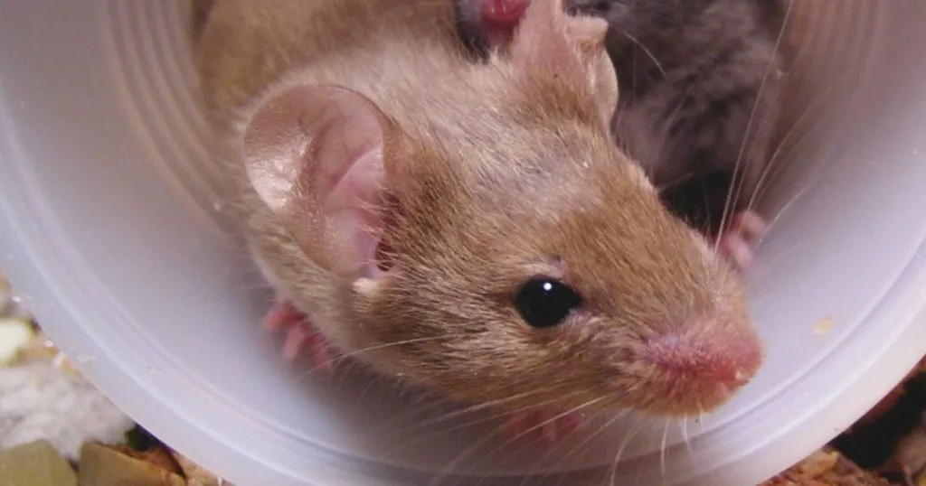
In this study, several behavioral tests were combined with techniques to stimulate, inhibit, and even monitor the activity of specific brain cells (see below). A first test consisted of animals freely exploring a two-chambered arena with unlimited access to food. Video tracking automatically monitored the whereabouts and movement of the animal. This method is easily combined with techniques such as optogenetics.
FREE TRIAL
Try EthoVision XT yourself!
Request a free trial and find out what EthoVision XT can do for your research!
-
check_circle
A cost-effective solution
-
check_circle
Powerful data selection
-
check_circle
Most cited video tracking system
Free Trial
arrow_forward
Mice also underwent real-time place preference tests in which one of the two chambers was paired with either optogenetic stimulation or inhibition, and self-stimulation tests in which the mouse received optogenetic stimulation (reward) upon nose-poking.
A PR3 (progressive ratio 3) test was used to measure motivation, and appetitive and consummatory behavior. After a nose poke, the mouse received a liquid caloric reward, then each successive reward required 3 more nose pokes than the previous. Licking while there was a reward (consummatory licks) or not (nonconsummatory licks) were measured.
Unraveling brain function at cell-level
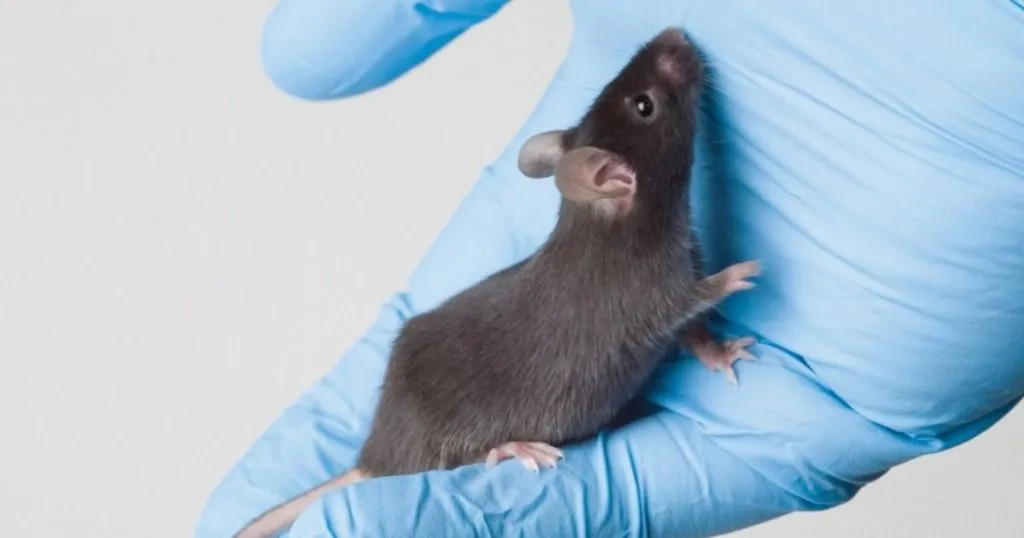
The specific brain cells Jennings and Ung focused on are a subset of LH neurons that produce GABA (gamma-Aminobutyric acid), an important inhibitory neurotransmitter in the mammalian nervous system. The researchers first explored functional role of these GABAergic LH neurons using optogenetics to selectively stimulate or inhibit the specific cells, combined with video tracking to automatically track the location and movement of the animals. (Read more about optogenetics in this blog post.)
When GABAergic neurons were stimulated, the mice spent more time in the food area and ate more. Mice also spent more time in an area that they had previously learned to associate with stimulation of these neurons. Optogenetic inhibition of these neurons led to less time spent in the food area, less eating, and less time in the location associated with this inhibition. The results implicate these neurons in the modulation of appetitive and consummatory behavior.
Confirming the essential role in appetitive response and consumption
The researchers used additional methods to further detail the role of the LH GABAergic neurons. Chemogenetic activation, for example, also enhanced consummatory behavior. But genetic ablation - essentially removing LH GABAergic neurons - blunted weight gain, reduced food intake, and decreased appetitive behavior and motivation, showing that these neurons are important for the regulation of appetitive responding, consumption, and energy balance.
Deep brain imaging to detect cell activity
So what remains is determining if appetitive and consummatory processes are regulated by the same or discrete neurons. To this end, the researchers used in vivo microendoscopic imaging, essentially implanting a mini-microscope into the mouse's skull. With this deep brain calcium imaging, the researchers were able to detect activity in individual LH GABAergic neurons while mice were freely moving and performing behavioral tasks.
Each cell has its own task
Researchers found that some neurons spiked in the presence of food, while others showed decreased activity. When mice had to work to get a reward, hundreds of neurons were active in response to either the consummation of the food reward (first lick after the reward delivery) or the unreinforced active nose pokes (appetitive response). Nonconsummatory licks after not receiving a reward for the nose poke led to much less neuronal activity.
Microendoscopic imaging exposed the functionally distinct subpopulations of LH GABAergic neurons that encode aspects of appetitive or consummatory behaviors, but rarely both. The same neurons that respond to the presence of food during the free access feeding task also responded to the consummation of the reward and the nose pokes during the PR3 task.
Mapping out the mosaic to find targets for addiction and eating disorders
With this research, Jennings, Ung, and their colleagues showed that the lateral hypothalamus has distinct neurons regulating either appetitive or consummatory behaviors. This suggests that separate networks in the brain regulate the motivation to eat and the actual consumption of the food.
The deep brain imaging technique in freely moving animals, especially during behavioral tests, has allowed the identification and even mapping of individual neurons. This no doubt opens up important opportunities for future research in specific targeting of these neurons and can eventually lead to the effective treatment of addictions and eating disorders.
References
Jennings, J.H.; Ung, R.L.; Resendez, S.L.; Stamatakis, A.M.; Taylor, J.G.; Huang, J.; Veleta, K.; Kantak, P.A.; Aita, M.; Shilling-Scrivo, K.; Ramakrishnan, C.; Deisseroth, K.; Otte, S.; Stuber, G.D. (2015). Visualizing hypothalamic network dynamics for appetitive and consummatory behaviors. Cell, 160, 516-527.
Be sure to check out the supplementary material, including impressive videos of the deep brain imaging and mouse behavior.